Muscle Tissues and Contractions
Part 1: Muscle Tissue and Contractions
Part 2: Types of Muscles
Part 3: Fast Twitch and Slow Twitch Muscles
Part 4: Physiological Effects of Strenuous Exercise
Muscle tissue is a contractive type of tissue that can undergo tension by shortening (concentric contractions), lengthening (eccentric contractions) or remaining the same length (isometric contractions).
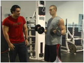
An example of a concentric contraction is performing the first part of a biceps curl where you hold a dumbbell down in one hand down by your side and then flex your elbow upward. The bicep muscle shortens in the process while undergoing tension. See the figures below:

Figure 1. Phases of a concentric contraction during a biceps curl.
First picture shows the start, second picture shows an intermediate position and the third shows the final position.
An example of an eccentric contraction is performing the second part of the biceps curl, where you slowly lower your hand back down to your side. The bicep muscle is still undergoing tension only this time it is lengthening. The same figures are shown below, just in reverse order.

Figure 2. Phases of an eccentric contraction during a biceps curl.
First picture shows the start, second picture shows an intermediate position and the third shows the final position.
An example of an isometric contraction is holding a bicep curl at a halfway point, or one where your forearm is parallel to the ground, for about 10 seconds. The length of the bicep muscle remains the same during this time period, however, it is still undergoing tension.

Figure 3. Isometric contraction.
This position is held for about 10 seconds.
The cells that make up your muscle tissue consist of the proteins actin and myosin. Each of these proteins is arranged into very fine threadlike filaments within the cell.
Myosin is a rather thick and active protein and is considered a molecular motor protein that is able to ratchet along the surface of a suitable substrate (actin). With each “turn of the ratchet”, they are able to grab the substrate and pull themselves in a given direction. Thus they are able to convert chemical energy into mechanical work. This is their main function.
Actin is a rather thin and passive protein when compared to myosin and has several functions within the cell. One of these functions is to give mechanical support to the cells and another is to allow for cell motility. With regards to muscle contraction or tension, actin serves as the substrate on which myosin proteins ratchet their force.
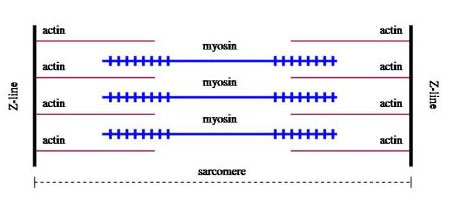
The basic unit of arrangement for the actin and myosin filaments within the cell is called a sarcomere. A sarcomere is arranged with the thick myosin filaments bordered by two actin filaments. See Figure 4.

Figure 4. Sarcomere before contraction.
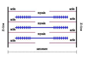
The ends of each thick myosin filament grab the adjacent thin actin filaments repeatedly, ratcheting and then letting go; the actin filaments are pulled closer together in the process and the sarcomere shortens during a concentric contraction.

Figure 5. Shortened sarcomere after contraction.
During an eccentric contraction, or one where the muscle lengthens while undergoing tension, this myosin filament detach from their actin filaments.
During an isometric contraction the myosin filaments hold firmly to the actin filaments without any movement.
Part 1: Muscle Tissue and Contractions
Part 2: Types of Muscles
Part 3: Fast Twitch and Slow Twitch Muscles
Part 4: Physiological Effects of Strenuous Exercise
[sc name=”2-stepoptin-blue”]
[sc name=”mma testimonial banner – green”]
[sc name=”Order Here Button-01″]


